Thoracic Muscles
These muscles can be divided into two groups: intrinsic and extrinsic.
- serratus posterior
- levatores costarum
- intercostal (external, internal, innermost)
- subcostal
- transversus thoracis
are primarily responsible for assisting with respiratory movements. They are often referred to as the respiratory muscles.
The extrinsic muscles of the thoracic wall consist of:
- subclavius
- pectoralis major and minor
- lower part of the serratus anterior muscle
Their primary function is to establish a functional connection between the thorax and the upper limb and neck. Through this connection, they assist in the movement of the shoulder (pectoral) girdle.
The pectoralis major is a large, fan-shaped muscle located in the chest. It covers the upper part of the chest and attaches to the humerus bone in the upper arm. The primary function of the pectoralis major is to bring the arm across the body (adduction) and to flex the arm at the shoulder joint.
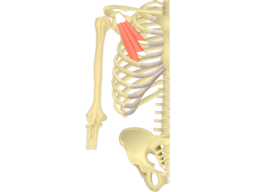
The pectoralis minor is a small, triangular muscle located in the upper chest, beneath the pectoralis major. The primary function of the pectoralis minor is to stabilize the scapula (shoulder blade) by drawing it forward and downward against the rib cage.
Serratus Anterior Muscle
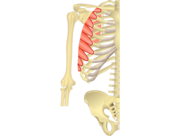
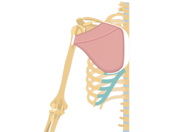
The serratus anterior is a muscle located on the lateral (side) surface of the ribcage. It extends from the upper eight or nine ribs to the scapula (shoulder blade). The primary function of the serratus anterior is to protract the scapula, which means pulling it forward and around the ribcage.
External Intercostal Muscles
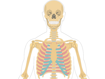
The external intercostal muscles are muscles located between the ribs. They run in an oblique direction from the lower border of one rib to the upper border of the rib below it. The primary function of the external intercostal muscles is to elevate the ribs during inhalation, which helps expand the chest and increase lung volume, allowing air to enter the lungs.
